


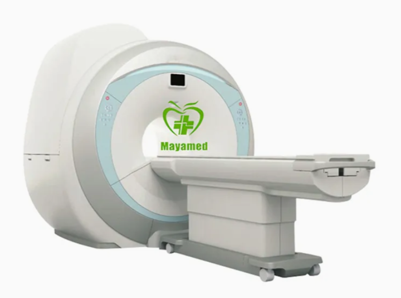
Low Field 0.35 T MRI Scanner
Low Field 0.35 T MRI Scanner
*Excellent price performance ratio
Comprehensive application suite and powerful software included in standard configuration
*Patient friendly appearance
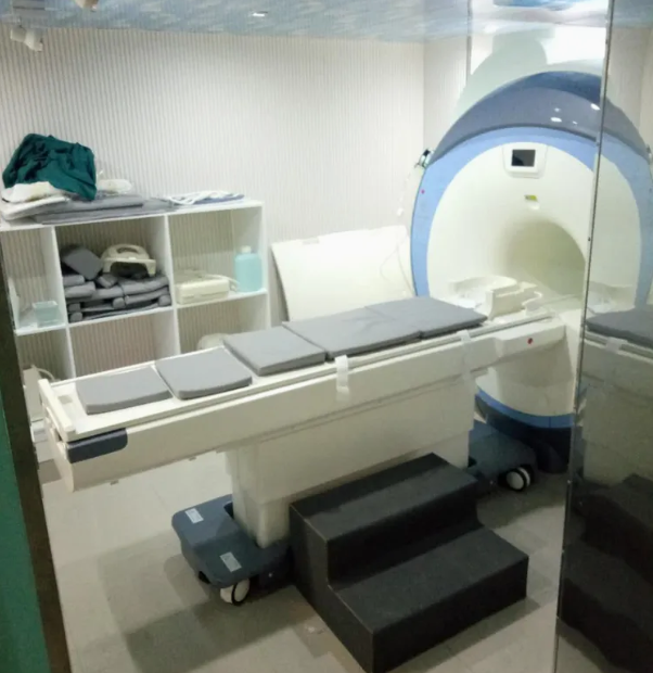
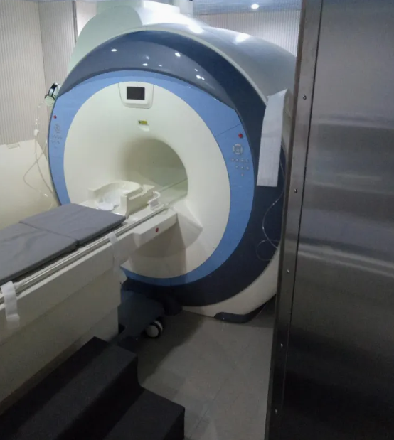
Most open C-shaped magnet
*Minimal sitting requirements
Less than 50 m² (538 sq.ft);
*Low operating Costs
Permanent magnet - no helium
*Higher revenues
Attractive to more patients and referring physicians
*Excellent Return On-Investment
Decreased costs-optimized profitability
*Efficient and Professional service team
24 hours service on-line
Specifications of MY-D054(0.35T) System
UUUUUUUUU1.0 Magnet:
Operating Field Strength 0.35 Tesla(3500 Gauss)
Magnet Type Full open C-shaped, 2-colum
Magnetic material Permanent Nd-Fe-B magnet
Homogeneity (400mm DSV) ≤ 2.0ppm (Vrms)
(200mm DSV) ≤ ±1ppm (FWHH)
Shimming Passive & active shimming
Patient gap 400mm(from cover to cover)
Net Weight 17,500KGS
Dimension 1970mm*1320mm*1820mm
UUUUUUUUU2.0 Gradient System:
Gradient strengh max. 25mT/m (Gx/Gy/Gz)
Slew rate 64mT/M/ms (Gx/Gy/Gz)
UUUUUUUUU3.0 RF System:
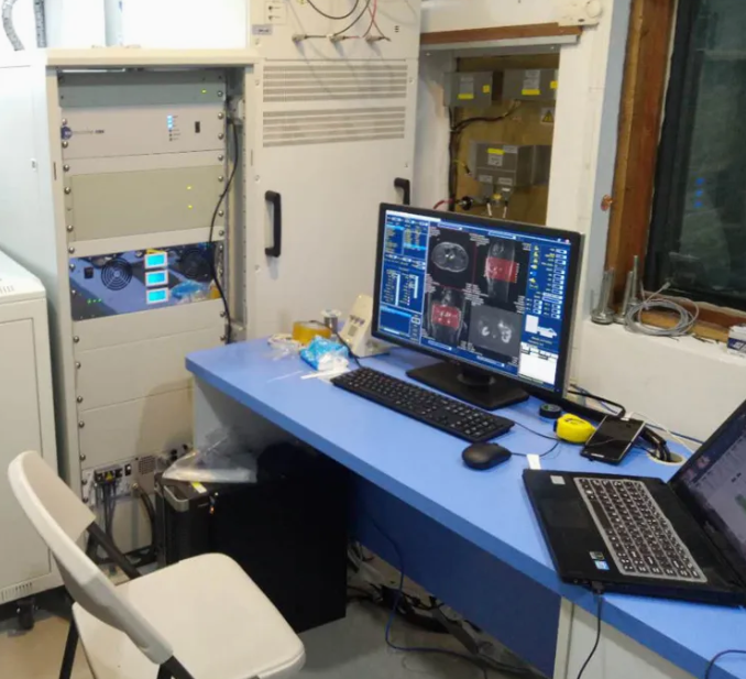
Spectrometer Digital (4 channels)
Transmitting coil Flat planar coil
Receiving coils Phase array coil (Neck coil, Head coil, Large body coil, Small body coil, Knee coil,)
Optional coils Wrist coil, Shoulder coil spine coil, breast coil, ankle coil, etc.
Power of transmitter amplifier 6KW
UUUUUUUUU4.0 Computer System:
Operating system Windows2000/NT/XP
CPU 2.8GHz(Dual Core Processor)
Hard disk ≥250GB
RAM 2048MB (2GB)
Display Device ≥22'' TFT
Network & laser printer interface DICOM3.0
Software BASDA BTI-035 Software
5.0 Auto Pre- scan
6.0 Pulse Sequence:
Standard IR
Spin Echo ( SE 2D/3D)
Multi-slice multi-echo (MSME)
Gradient Echo (GRE 2D/3D)
Steady state process gradient echo( SSPGRE)
Fast spin echo (FSE)
Single shot fast spin echo(SSFSE)
Multi shot fast spin echo (MSFSE)
Inversion recovery (IR)
Inversion recovery fast spin echo (IRFSE)
Short time inversion recovery (STIR)
Fluid attenuated inversion recovery ( FLAIR)
MRM ,MRU ,MRCP
TOF 2D/3D MRA
Diffusion weighted imaging (DWI)
Echo planar imaging(EPI)
7.0 Image:
Acquisition matrix 64/128/256/512
Resolution 1mm(head 24cm FOV 256X256)
1.5MM(body 30cm FOV 256X256)
0.5mm(head 24cm FOV 512X512)
0.75mm( body 30cm FOV 512X512)
Maximum display matrix 1024x1024
FOV 20 ~ 400mm
Slice Orientation Sagittal, coronal, transversal, any angle any oblique
Image Type T1 Weighted imaging , T2 weighted imaging ,T2* weighted imaging , proton density imaging;Water Suppressed Imange,Fat Suppressed Image, MRM, MRU,MRCP; Magnetic Resonance angiography (MRA), Diffusion Weighted Imaging (DWI)
8.0 Patient Table:
Max. Patient load about 240kg;
Available with laser light localizer for patient positioning
Longitudinal travel range: ≥1650mm
Intercommunication between patient and operator: available
9.0 Power Supply
3N~380/400V 50/60Hz 10KVA
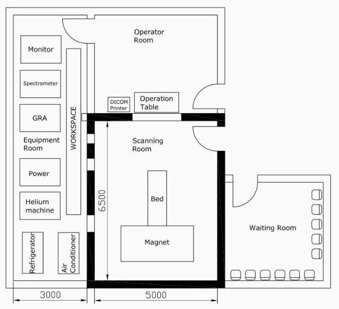
10.0 Typical Layout:
Magnet Room About 30 Square meters(5m×6m);
Equipment Room About 9 Square meters(3m×3m);
Control Room About 9 Square meters(3m×3m);
Total MRI-System area About 48 Square meters.
1. Magnet System
| Feature | Specification | Analysis |
|---|---|---|
| Field Strength | 0.35 Tesla (3500 Gauss) | This is a lower magnetic field strength compared to higher-end models like 1.5T or 3T systems. While it may be suitable for basic imaging, it limits the resolution and speed of imaging, making it less ideal for detailed brain, musculoskeletal, or cardiac imaging. |
| Magnet Type | Full open C-shaped, 2-column | C-shaped design maximizes patient comfort by offering greater access and reducing the feeling of confinement. This can increase patient acceptance and may be beneficial for claustrophobic patients. |
| Magnetic Material | Permanent Nd-Fe-B magnet | Permanent magnets eliminate the need for costly helium and cooling systems, significantly reducing operational costs. However, the lower field strength may limit imaging capabilities. |
| Shimming | Passive & Active Shimming | Dual shimming (passive and active) helps maintain uniformity in the magnetic field, improving image quality and reducing distortion. |
| Homogeneity | ≤ 2.0ppm (400mm DSV), ≤ ±1ppm (200mm DSV) | Acceptable homogeneity for a 0.35T system, but lower than what you would expect from 1.5T or higher systems. This could affect the quality of certain high-resolution imaging. |
| Patient Gap | 400mm | The gap allows for comfortable patient positioning and accessibility, making it better for accommodating larger patients compared to standard narrow-bore designs. |
2. Gradient System
| Feature | Specification | Analysis |
|---|---|---|
| Gradient Strength | 25mT/m (Gx/Gy/Gz) | A lower gradient strength compared to higher-end MRI machines. While it is sufficient for basic imaging, it may struggle with more advanced techniques like diffusion tensor imaging (DTI) or functional MRI (fMRI). |
| Slew Rate | 64mT/m/ms | This is a decent slew rate for general imaging. However, faster imaging techniques such as echo planar imaging (EPI) or cardiac imaging may be limited in speed and accuracy. |
3. RF System
| Feature | Specification | Analysis |
|---|---|---|
| Spectrometer | Digital (4 channels) | Digital spectrometers offer better signal acquisition and faster processing. However, with 4 channels, this may limit the flexibility of more advanced multi-slice imaging or complex scans. |
| Transmitting Coil | Flat planar coil | Offers decent coverage for body imaging but may be limited for small structures like the brain or joints. |
| Receiving Coils | Phase array coil (Neck, Head, Body, Knee, etc.) | A variety of coils provide versatility for different imaging needs, improving patient comfort by targeting specific regions with minimal repositioning. |
| Power of Amplifier | 6KW | This is relatively modest power, but adequate for a 0.35T system. However, high-power sequences may have reduced efficiency. |
4. Computer System
| Feature | Specification | Analysis |
|---|---|---|
| Operating System | Windows 2000/NT/XP | Outdated OS, which may lead to security and compatibility issues. A more modern OS such as Windows 10 or embedded Linux would be ideal for a better user experience and system longevity. |
| CPU | 2.8GHz (Dual Core) | Decent performance for general use but may struggle with intensive image reconstruction. |
| Hard Disk | ≥250GB | Sufficient for storing moderate data, though an upgrade to larger capacity may be needed depending on the volume of imaging data. |
| RAM | 2GB | Minimal RAM — while functional, a higher RAM capacity would ensure faster processing and enhanced system performance, especially for larger datasets. |
| Display | ≥22'' TFT | Acceptable for basic image display but would benefit from higher resolution and larger screens for detailed interpretation of complex images. |
| Software | BASDA BTI-035 Software | Proprietary software should cover basic MRI needs but may lack advanced processing options available with higher-end systems. |
5. Pulse Sequences
| Feature | Specification | Analysis |
|---|---|---|
| Pulse Sequences | Includes a wide variety (Spin Echo, Gradient Echo, FSE, Diffusion, MRA, DWI, etc.) | Offers comprehensive sequence options for general clinical and research applications. While useful for basic scans, advanced sequences like fMRI or cardiac MRI may not perform as well with this system. |
6. Image Quality
| Feature | Specification | Analysis |
|---|---|---|
| Matrix | 64/128/256/512 | The available matrix sizes will allow reasonable resolution for general imaging. Higher matrix sizes are available but may strain the system’s performance, especially at 0.35T. |
| Resolution | 1mm (head), 1.5mm (body) | Adequate for basic clinical applications. High-resolution scans, especially for smaller structures, may not be as crisp as those from higher-field MRI systems (1.5T and above). |
| Maximum Display Matrix | 1024x1024 | A higher display matrix would ensure the best possible image clarity, but limitations in field strength and gradient performance may still lead to suboptimal results in some cases. |
| FOV (Field of View) | 20~400mm | Flexible, but limited compared to higher-field systems in terms of deeper tissue penetration and resolution. |
7. Patient Table & Comfort
| Feature | Specification | Analysis |
|---|---|---|
| Max Patient Load | 240kg | Suitable for most patients, including larger individuals. |
| Table Travel | ≥1650mm | Provides enough space for patient positioning and accommodates a wide variety of patient sizes. |
| Intercommunication | Available | Ensures effective communication between the operator and patient during the scan. |
8. Power Supply & Layout
| Feature | Specification | Analysis |
|---|---|---|
| Power Supply | 3N~380/400V 50/60Hz 10KVA | Requires a stable power source, but within typical MRI system power requirements. |
| Room Layout | ~48m² | Compact, requiring minimal space for MRI installation. This makes it suitable for smaller facilities or locations where space is limited. |
9. Cost-Effectiveness & ROI
| Feature | Specification | Analysis |
|---|---|---|
| Price-Performance Ratio | Excellent | With a low operating cost (no helium needed), this MRI system can be a cost-effective solution for clinics or smaller hospitals. It’s a solid option for facilities that need basic diagnostic capabilities but don’t have the budget for high-field MRI systems. |
| Patient Attraction | Yes | Its open design, lower price, and reduced operational costs make it an attractive option for a wide range of patients and referring physicians, potentially increasing patient volume and revenues. |
| Return on Investment (ROI) | High | Low maintenance and operational costs (no helium or liquid nitrogen) can significantly improve profitability. The system is ideal for high patient throughput with a smaller initial investment. |
Overall Conclusion
The MY-D054 (0.35T) MRI system is an affordable, low-maintenance option that excels in cost-efficiency and patient comfort, making it ideal for smaller clinics or hospitals. However, its lower field strength (0.35T) means that it is not suitable for high-resolution, detailed imaging like brain, musculoskeletal, or cardiac scans. Its open design and low operational costs (no helium) make it appealing for patient comfort and ongoing cost savings.
For facilities focused on general imaging, patient accessibility, and cost reduction, this system provides a solid, cost-effective solution. However, those requiring advanced imaging capabilities or high-resolution scans may need to consider higher field strength systems (e.g., 1.5T or 3T).
Share
Total des articles
Sous-total des produits