


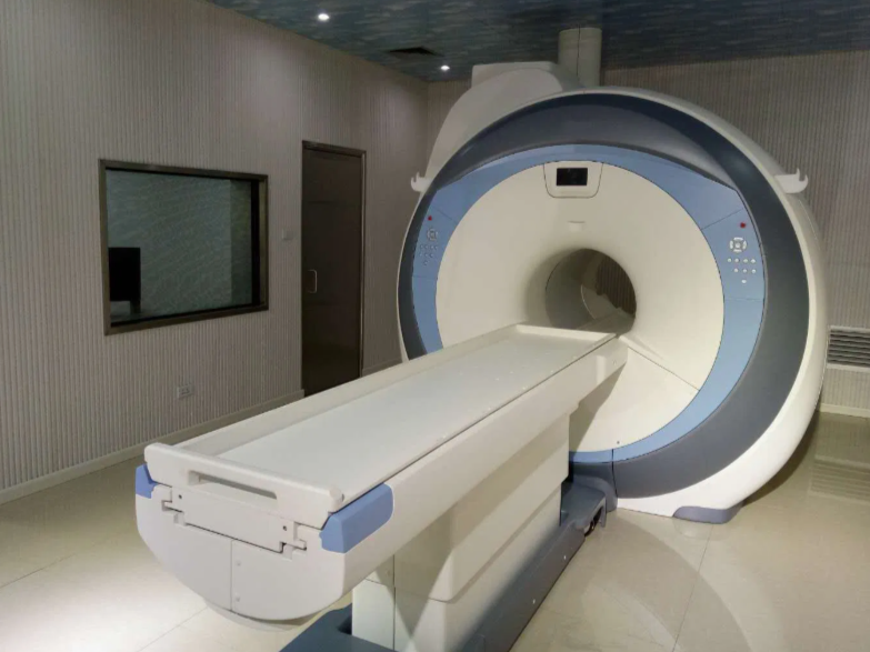
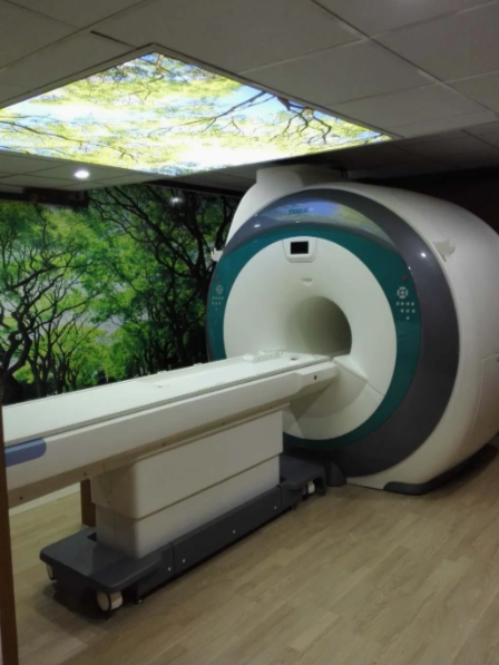
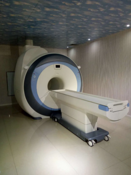
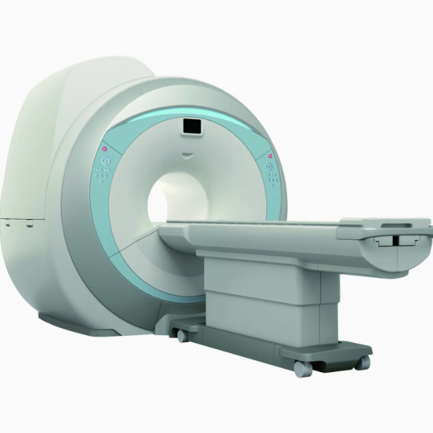
1.5t MRI Superconducting MRI Scanner (CE & ISO)
1.5t MRI Superconducting MRI Scanner (CE & ISO)
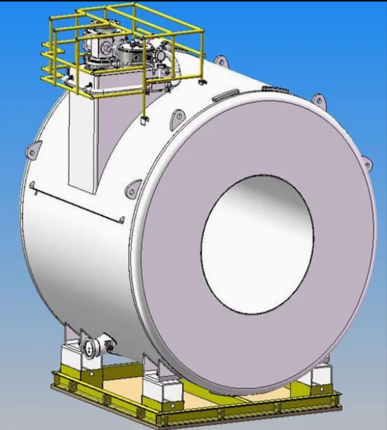
1.5T active shield superconducting magnet
Gradient System (Performance Controls Incorporation, USA)
RF System: Spectrometer: All-digital (MR Solutions Ltd., UK)
RF Amplifier: Peak power:18KW (Analogic, USA)
Transmitting Coil
Computer system: CPU: dual core ≥2.8GHz; RAM: ≥2GB; Hard disk: ≥500GB Main display: ≥24´´LCD TFT monitoring
Console: New communication user software interface, bilingual choice Operation system: Windows XP Professional Monitoring devices, machine monitoring, patient monitoring, multi-parameter monitoring
Ref: MKT-20160815
No. Item Specification
1 Magnet System
1.1 Magnet Type Superconductive
1.2 Field Strength 1.5T
1.3 Shielding Method Active
1.4 Shimming Method and Type Active + Passive + Dynamic
1.5 Magnet stability ≤0.1ppm/h
1.6 Homogeneity (DSV, VRMS)
1.6.1 45cm ≤0.832ppm
1.6.2 40cm ≤0.323ppm
1.6.3 30cm ≤0.107ppm
1.6.4 20cm ≤0.041ppm
1.6.5 10cm ≤0.01ppm
1.7 Length of magnet(exclude cover) 157cm
1.8 Inner diameter of magnet 600mm
1.9 5 Gauss fringe field(X,Y,Z axis) ≤2.5m x 2.5m x 4.0m
Maximum gradient field (single axis, invalid) 41mT/m
2.2 Maximum gradient slew rate(single axis,
invalid)
187mT/m/ms
2.3 Minimum gradient rise time 0.22ms
2.4 Maximum gradient field and slew rate reached
at the same time
Yes
2.5 Gradient cooling system Water cooling
2.6 Full digital real-time transmit and receiving
gradient control system
Yes
3 RF System
3.1 Max RF amplifier power 18kW
3.2 Center frequency 63.87MHz
3.3 Real-time digital RF energy monitoring Yes
3.4
Real-time digital RF short-term accumulation
monitoring
Yes
3.5 Real-time digital RF long-term accumulation Yes
1. Magnet System
| Feature | Specification | Analysis |
|---|---|---|
| Type | Superconductive | Gold standard in modern MRI. Superconductive magnets ensure stable and powerful magnetic fields essential for high-quality imaging. |
| Field Strength | 1.5 Tesla | Commonly used clinical strength, offering a balance between image quality and patient safety. Sufficient for neuro, musculoskeletal, abdominal, and cardiovascular imaging. |
| Shielding | Active | Reduces magnetic fringe fields, minimizing infrastructure costs (e.g., less room shielding required). Enhances safety in nearby environments. |
| Shimming | Active + Passive + Dynamic | Ensures excellent field homogeneity, leading to clearer images with fewer artifacts. Multiple shimming modes allow adaptation to different imaging situations. |
| Stability | ≤ 0.1 ppm/h | Highly stable — ensures that image quality remains consistent over time. |
| Homogeneity (DSV/VRMS) | From ≤0.832ppm at 45cm to ≤0.01ppm at 10cm | Very high homogeneity. Ideal for both small region-of-interest scans and whole-body imaging. |
| Inner Bore | 600 mm | Standard patient bore. Comfortable for most patients but not ultra-wide. |
| Length | 157 cm (excluding cover) | Reasonably compact for a superconducting magnet. May allow more flexible room layouts. |
| 5 Gauss Line | ≤2.5m x 2.5m x 4.0m | Compact fringe field helps ensure safety zones are manageable — critical for MRI room planning. |
2. Gradient System (Performance Controls Inc., USA)
| Feature | Specification | Analysis |
|---|---|---|
| Max Gradient Field | 41 mT/m | Adequate for clinical and advanced imaging sequences (e.g., DWI, fMRI). |
| Max Slew Rate | 187 mT/m/ms | Excellent — enables fast imaging, essential for dynamic studies, cardiac imaging, etc. |
| Minimum Rise Time | 0.22 ms | Fast — supports high temporal resolution imaging. |
| Simultaneous Max Gradient & Slew | Yes | Indicates well-optimized hardware performance. |
| Cooling | Water Cooling | Ensures consistent performance and prevents overheating, especially during demanding sequences. |
| Control System | Full digital real-time | Enhances synchronization and reduces distortion/artifacts. Modern and efficient. |
3. RF System (MR Solutions Ltd. & Analogic, USA)
| Feature | Specification | Analysis |
|---|---|---|
| Spectrometer | All-digital | Modern and reliable, allows precise signal acquisition. |
| Amplifier Power | 18kW | High power ensures deep tissue penetration and high-quality signal-to-noise ratios. |
| Center Frequency | 63.87 MHz | Appropriate for 1.5T field strength — ensures resonance of hydrogen protons. |
| Digital Monitoring | Real-time monitoring (energy + accumulation, short & long term) | Improves patient safety, system protection, and data fidelity. Reflects an advanced and safety-focused design. |
| Transmitting Coil | Included | Ensures compatibility and optimized performance. Likely a body or volume coil for transmission. |
4. Computer System & Console
| Feature | Specification | Analysis |
|---|---|---|
| CPU | Dual Core ≥2.8GHz | Decent, but may be slightly outdated by today’s high-end imaging workstations. Can still support clinical operations. |
| RAM | ≥2GB | Low by modern standards. Recommend upgrade to 8GB+ for faster reconstruction and processing. |
| Display | ≥24" LCD TFT | Acceptable; large enough for viewing high-res scans. |
| Console Software | Bilingual, user-friendly | Promotes accessibility and user efficiency. |
| Operating System | Windows XP Professional | Outdated — may pose cybersecurity risks. Strongly recommend update to Windows 10 or embedded Linux for stability and support. |
5. Monitoring Systems
| Feature | Specification | Analysis |
|---|---|---|
| Machine & Patient Monitoring | Multi-parameter | Vital for patient safety and clinical use, especially in anesthetized or critical care patients. |
✅ Strengths:
-
Industry-standard 1.5T with excellent homogeneity and stability
-
High-end gradient and RF specs for diverse clinical use
-
Fully digital systems with safety-focused monitoring
-
Compact, active-shielded design for flexible installation
⚠️ Considerations / Recommendations:
-
Consider a wide-bore version (70cm) if patient comfort or obesity cases are a concern
-
Ensure proper cooling infrastructure for the water-cooled gradient system
-
Verify availability of local service support or training for longevity
Share
Total des articles
Sous-total des produits